Services
Ultrasound guided procedures:
Ultrasound-guided procedures refer to medical interventions or treatments that are performed with the assistance of ultrasound imaging technology. Ultrasound imaging uses high-frequency sound waves to create real-time images of the body’s internal structures, allowing healthcare providers to visualize and guide instruments with precision. Ultrasound guidance enhances the accuracy and safety of various medical procedures, including but not limited to:
- Joint Aspiration and Injection: As described earlier, ultrasound guidance can aid in the accurate placement of a needle into a joint space for the aspiration of fluid or injection of medication.
- Biopsy: Ultrasound-guided biopsies involve the sampling of tissue or cells from a specific organ or area of concern under ultrasound visualization. This technique is commonly used to diagnose conditions such as tumors, cysts, or infections.
- Nerve Blocks: Ultrasound-guided nerve blocks involve the precise delivery of local anesthetic medication to target specific nerves responsible for pain sensation. This approach is often used for pain management in conditions such as chronic pain, nerve injuries, or regional anesthesia for surgical procedures.
- Vascular Access: Ultrasound guidance is used to identify and access veins or arteries for procedures such as central venous catheter placement, peripheral intravenous line insertion, or arterial puncture for blood gas analysis.
- Soft Tissue Injections: Ultrasound imaging can assist in the accurate placement of injections into soft tissues, such as tendons, ligaments, muscles, or bursae, for the treatment of conditions like tendonitis, bursitis, or muscle strains.
- Fluid Drainage: Ultrasound-guided drainage procedures involve the removal of fluid collections, such as abscesses or seromas, under real-time ultrasound visualization to ensure precise needle placement and effective drainage.
- Aspiration of Cysts: Ultrasound guidance can aid in the aspiration or drainage of cysts, such as ovarian cysts or cystic lesions in the breast, kidneys, or liver, to relieve symptoms and assess the fluid content for diagnostic purposes.
- Injection of Therapeutic Agents: Ultrasound imaging can assist in the delivery of therapeutic agents, such as platelet-rich plasma (PRP), stem cells, or prolotherapy solutions, into target tissues for regenerative or anti-inflammatory purposes.
- Ultrasound-guided procedures offer several advantages, including improved accuracy, reduced risk of complications, real-time visualization of anatomical structures, and enhanced patient comfort. These procedures are typically performed by trained healthcare providers, including radiologists, interventional radiologists, anesthesiologists, or other specialists with expertise in ultrasound-guided interventions.
Ultrasound-guided joint aspirations:
Also known as ultrasound-guided joint aspirations and injections, involve the use of ultrasound imaging to guide the placement of a needle into a joint space for the purpose of removing fluid (aspiration) or injecting medication (injection). This procedure is commonly performed to diagnose and treat various joint-related conditions, including arthritis, bursitis, and joint effusions. The patient is positioned comfortably, typically lying down or seated, with the joint to be aspirated or injected exposed and accessible. The skin overlying the joint is cleaned and sterilized to reduce the risk of infection.
A small amount of gel is applied to the skin over the joint, and an ultrasound probe is placed on the gel-covered area. The ultrasound machine emits high-frequency sound waves that create real-time images of the joint and surrounding structures on a monitor. Using the ultrasound images as a guide, the healthcare provider identifies the optimal location for needle insertion into the joint space. The needle is carefully advanced under continuous ultrasound guidance to ensure precise placement within the joint.
Once the needle is correctly positioned within the joint space, fluid is withdrawn from the joint using a syringe attached to the needle. This fluid can be sent to a laboratory for analysis to help diagnose the underlying cause of joint inflammation or swelling. Ultrasound-guided joint aspirations and injections offer several advantages over traditional blind techniques, including improved accuracy, reduced risk of complications, and real-time visualization of needle placement. This procedure is generally safe and well-tolerated by patients, with minimal discomfort and a low risk of adverse effects when performed by trained healthcare professionals. However, as with any medical procedure, there are potential risks and contraindications that should be discussed with your healthcare provider before undergoing ultrasound-guided joint aspirations.
Ultrasound-Guided Corticosteroid Injections
Once the needle is in the correct position, the corticosteroid medication, often mixed with a local anesthetic for immediate pain relief, is injected into the affected area. The medication helps reduce inflammation, alleviate pain, and promote healing of the underlying condition. After the injection, the patient may be advised to rest the treated area for a short period. Some patients may experience temporary soreness or swelling at the injection site, which typically resolves within a few days. Patients are usually instructed to resume normal activities gradually, as tolerated.
Ultrasound-guided corticosteroid injections offer several advantages over traditional blind injections, including: Increased Accuracy: Ultrasound guidance allows for precise needle placement, ensuring that the medication is delivered directly to the affected area.
Real-Time Visualization: Healthcare providers can visualize the needle in real-time as it approaches the target site, reducing the risk of inadvertent injury to surrounding structures.
Improved Outcomes: By delivering medication directly to the site of inflammation or injury, ultrasound-guided injections may lead to better treatment outcomes and faster symptom relief compared to blind injections.
Overall, ultrasound-guided corticosteroid injections are a safe and effective treatment option for managing musculoskeletal pain and inflammation, with the potential to improve patient outcomes and quality of life.
Ultrasound-Guided Viscosupplementation/Hyaluronic Acid Joint Injections:
Ultrasound-guided viscosupplementation is a procedure used to treat osteoarthritis, particularly in weight-bearing joints such as the knee. It involves the injection of a thick fluid called hyaluronic acid (HA) directly into the joint space under the guidance of ultrasound imaging technology. Here’s an overview of the procedure:
- Patient Evaluation: Before the procedure, the patient undergoes a thorough evaluation, which may include a physical examination, review of medical history, and diagnostic imaging studies such as X-rays or MRI scans. This helps determine if viscosupplementation is an appropriate treatment option.
- Patient Preparation: On the day of the procedure, the patient is positioned comfortably, usually lying down or sitting, with the affected joint exposed and accessible. The skin over the injection site is cleaned and sterilized to minimize the risk of infection.
- Ultrasound Imaging: A healthcare provider trained in ultrasound-guided procedures uses an ultrasound machine to visualize the joint in real-time. Ultrasound imaging allows the provider to accurately identify the joint space, assess the integrity of surrounding structures, and locate areas of inflammation or damage.
- Needle Placement: Using the ultrasound images as a guide, the provider inserts a thin needle through the skin and into the joint space. The needle is advanced until its tip is positioned accurately within the joint capsule.
- Injection: Once the needle is in the correct position, the viscosupplement, typically hyaluronic acid, is injected into the joint space. Hyaluronic acid is a naturally occurring substance found in the synovial fluid of joints, where it acts as a lubricant and shock absorber. By supplementing the natural HA in the joint, viscosupplementation aims to improve joint lubrication, reduce friction, and alleviate pain associated with osteoarthritis.
- Post-Injection Care: After the injection, the patient may be advised to rest the treated joint for a short period. Some patients may experience mild discomfort or swelling at the injection site, which usually resolves within a few days. Patients are typically instructed to avoid strenuous activities for a brief period following the procedure.
Ultrasound-guided viscosupplementation offers several advantages over blind injections, including improved accuracy, real-time visualization of the injection site, and reduced risk of complications. It is generally well-tolerated and may provide symptomatic relief for patients with osteoarthritis, particularly those who have not responded to conservative treatments such as oral medications, physical therapy, or corticosteroid injections. As with any medical procedure, the potential risks and benefits of viscosupplementation should be discussed with a qualified healthcare provider.
Ultrasound-Guided Platelet Rich Plasma (PRP) Injections
Ultrasound-guided platelet-rich plasma (PRP) injections are a minimally invasive medical procedure used to treat various musculoskeletal conditions, such as tendon injuries, ligament injuries, osteoarthritis, and other soft tissue injuries. Here’s an overview of the procedure:
Prior to the Injection:
- NSAID Avoidance: Refrain from taking NSAIDs (Nonsteroidal Anti-Inflammatory Drugs) such as Motrin, Aleve, Ibuprofen, or Advil for two weeks before the procedure.
- Hydration: Increase fluid intake 24 hours before the procedure to ensure adequate hydration.
- Steroid Injection: Avoid receiving steroid injections within the past 2-3 weeks before the PRP
Patient Evaluation: Before the procedure, the patient undergoes a comprehensive evaluation by a healthcare provider, which may include a physical examination, review of medical history, and diagnostic imaging studies such as ultrasound, MRI, or X-rays. This evaluation helps determine if PRP therapy is an appropriate treatment option for the patient’s condition.
Preparation of PRP: Prior to the injection, a small sample of the patient’s blood is collected through a standard blood draw procedure. The blood sample is then processed using a centrifuge machine to separate the platelets from other components of the blood, such as red blood cells and plasma. This process concentrates the platelets, which contain growth factors and other bioactive proteins that play a key role in tissue repair and regeneration.
Ultrasound Imaging: During the procedure, the patient is positioned comfortably, and the affected area is exposed and accessible. A healthcare provider trained in ultrasound-guided procedures uses an ultrasound machine to visualize the targeted area in real-time. Ultrasound imaging allows the provider to accurately identify the specific anatomical structures, such as tendons, ligaments, or joints, where the PRP injection will be administered.
Needle Placement: Using the ultrasound images as a guide, the provider inserts a thin needle into the targeted area. The needle is carefully positioned to ensure precise delivery of the PRP into the affected tissue or joint space.
Injection of PRP: Once the needle is in the correct position, the concentrated PRP is injected into the targeted area. The growth factors and bioactive proteins present in the PRP solution stimulate the body’s natural healing processes, promoting tissue repair, reducing inflammation, and relieving pain.
After the Injection:
- Expected Discomfort: Expect discomfort or pain, particularly in the first 20-30 minutes after the procedure. Soreness or mild pain may persist for about one week afterward, varying depending on the patient and the injection site.
- Management: Apply ice to the injection site to help alleviate pain and reduce swelling.
- Medication Restrictions: Avoid NSAIDs for the first 4 weeks after the procedure. Instead, you can take Tylenol or other pain medications for short-term relief during the first week.
- Activity Restrictions:
- Lower Extremity Injections: Refrain from high-impact activities like running, jumping, or spinning for 2-4 weeks.
- Upper Extremity Injections: Avoid heavy lifting and excessive upper extremity activities for 2-4 weeks.
- Follow-up: Schedule a follow-up appointment with your doctor, usually 2-4 weeks after the injection. During this visit, the doctor will assess your progress and determine if you can gradually resume normal activities or if physical therapy is necessary.
Risks:
Infection: There is a low risk of infection whenever the skin is penetrated, but proper sterile techniques minimize this risk.
PRP Risks: Since PRP is derived from your own blood, the risks associated with it are minimal.
Injection Site Risks:
Skin Blanching: Rarely, the skin may turn white at the injection site.
Skin Dimpling: In some cases, the skin may dimple.
Fat Necrosis/Skin Atrophy: This rare complication may lead to skin changes or fat tissue damage at the injection site, which could take up to a year or more to resolve.
It’s essential to discuss any concerns or questions you have with your healthcare provider before undergoing the procedure. Your provider will provide personalized guidance and ensure that you receive optimal care throughout the treatment process.
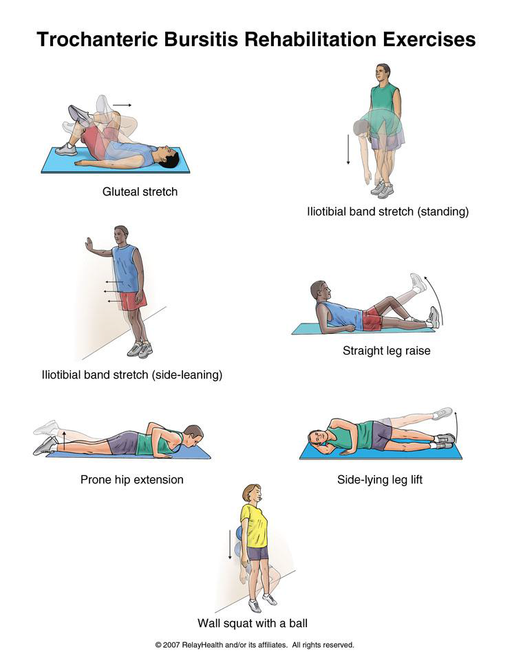
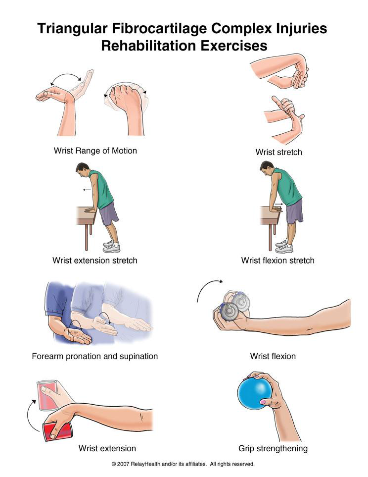
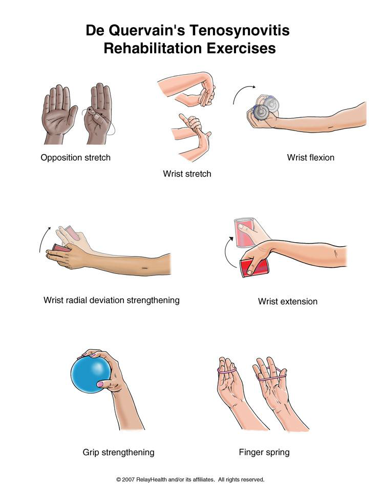
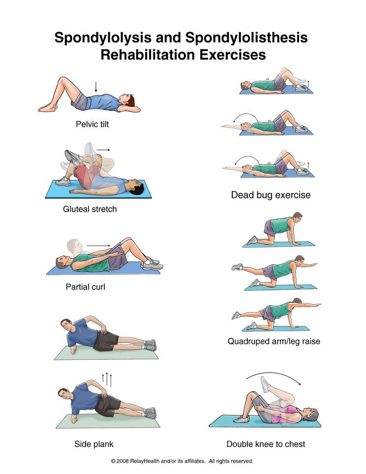
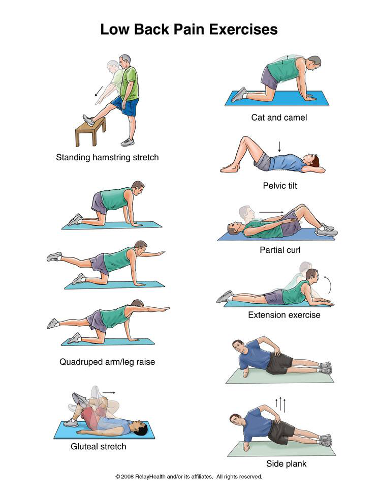
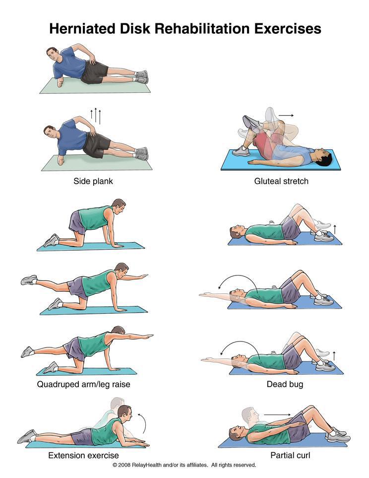
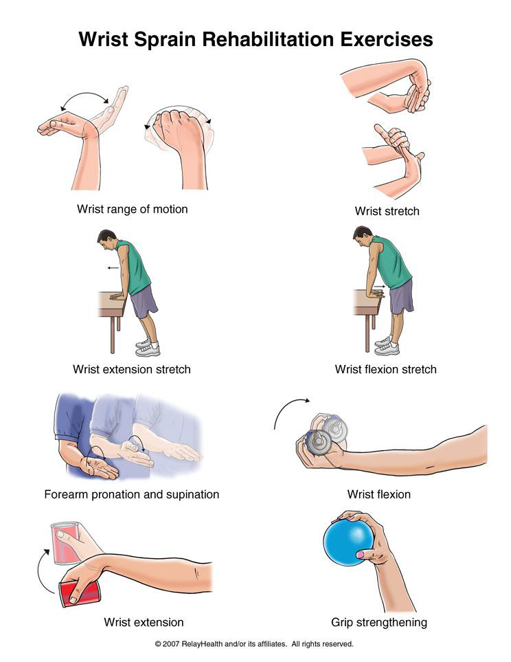
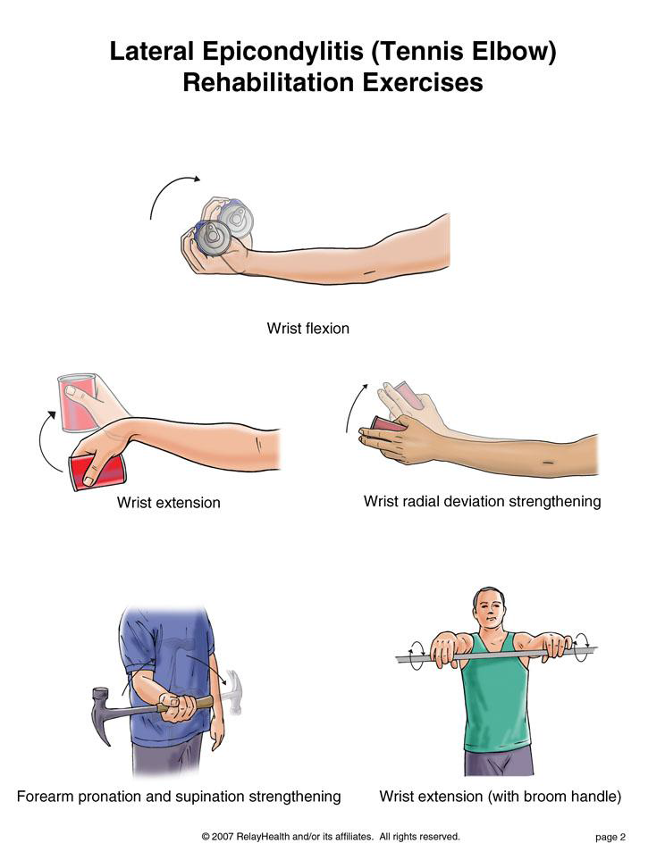
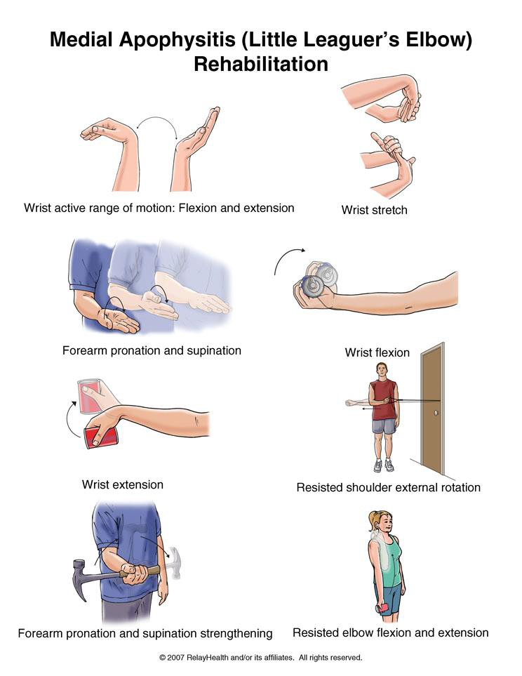
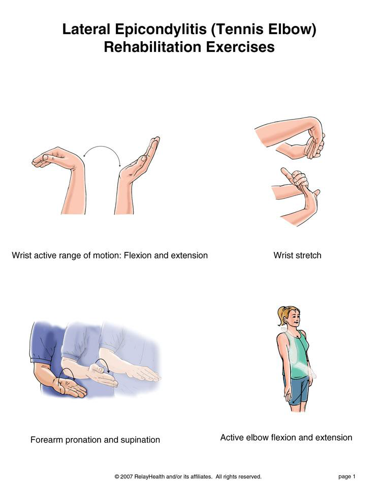
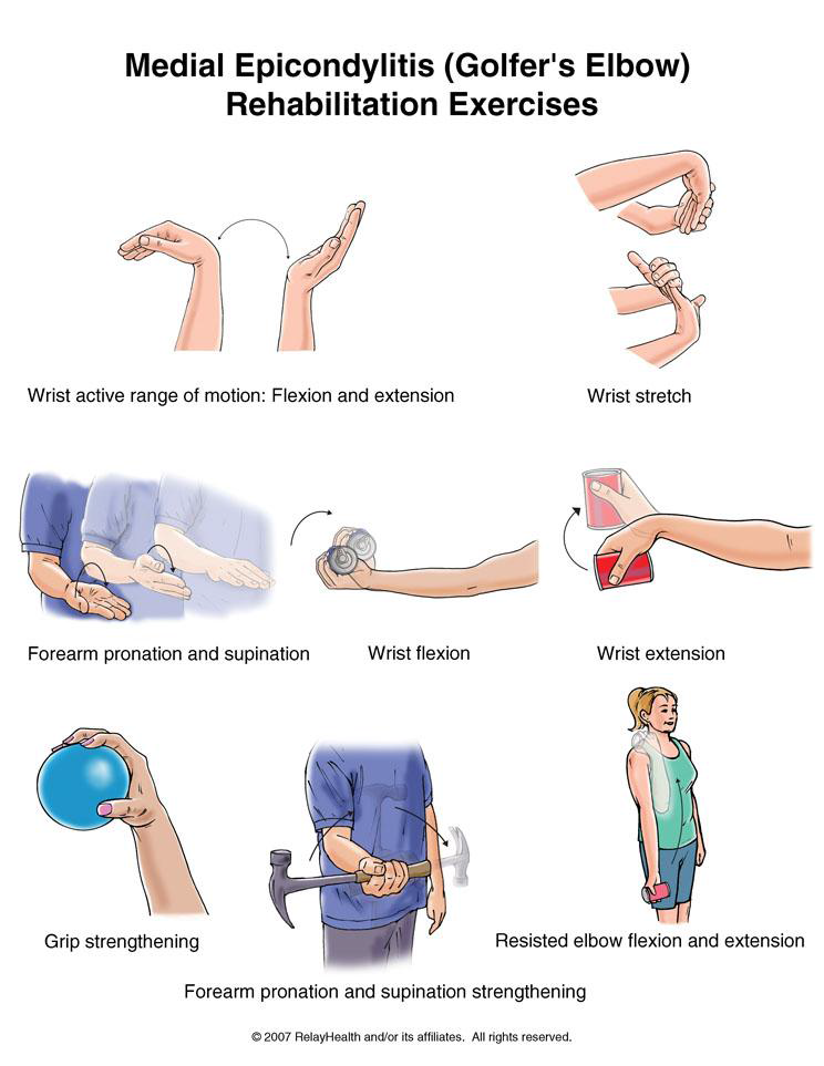
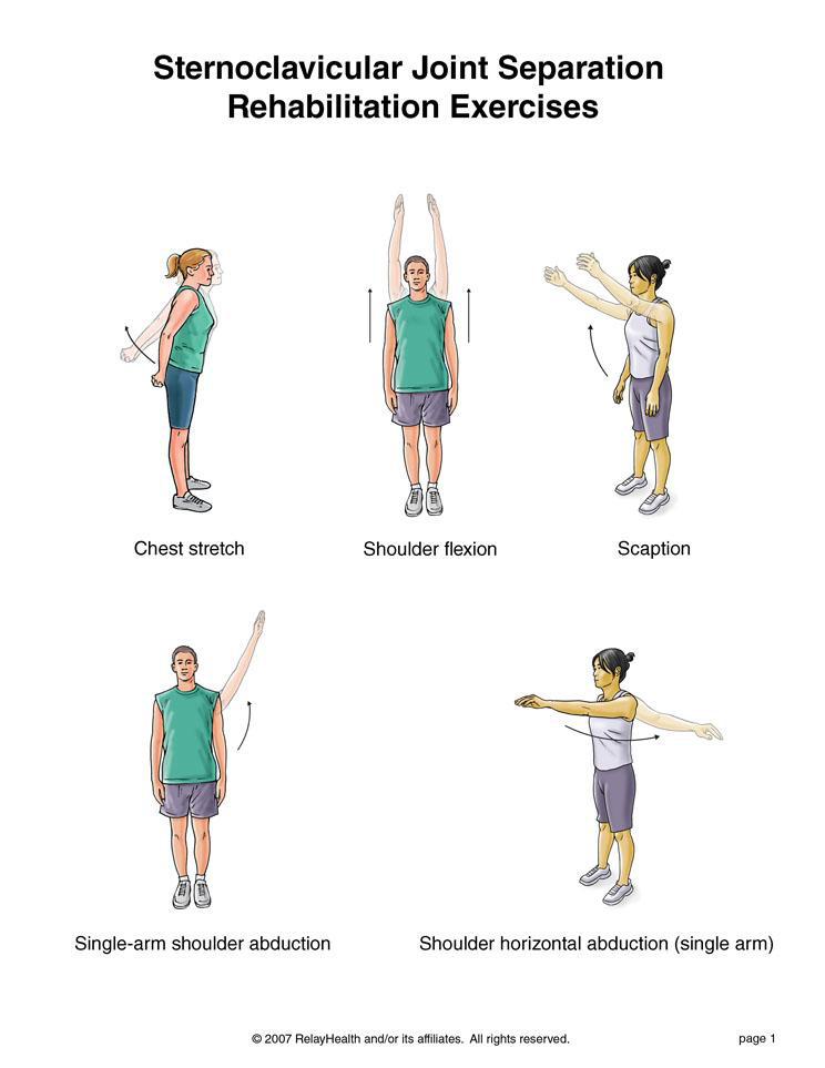
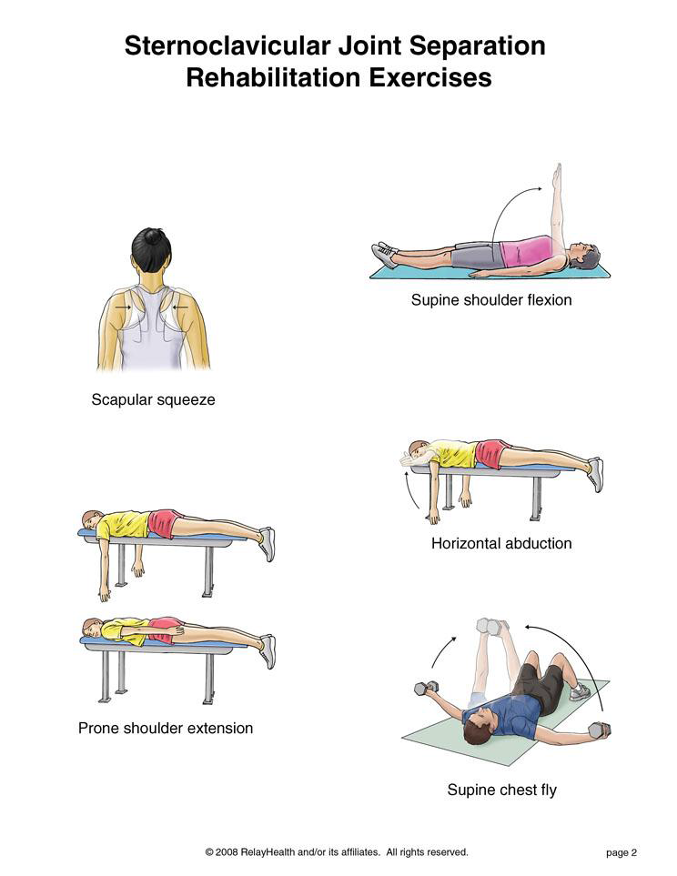
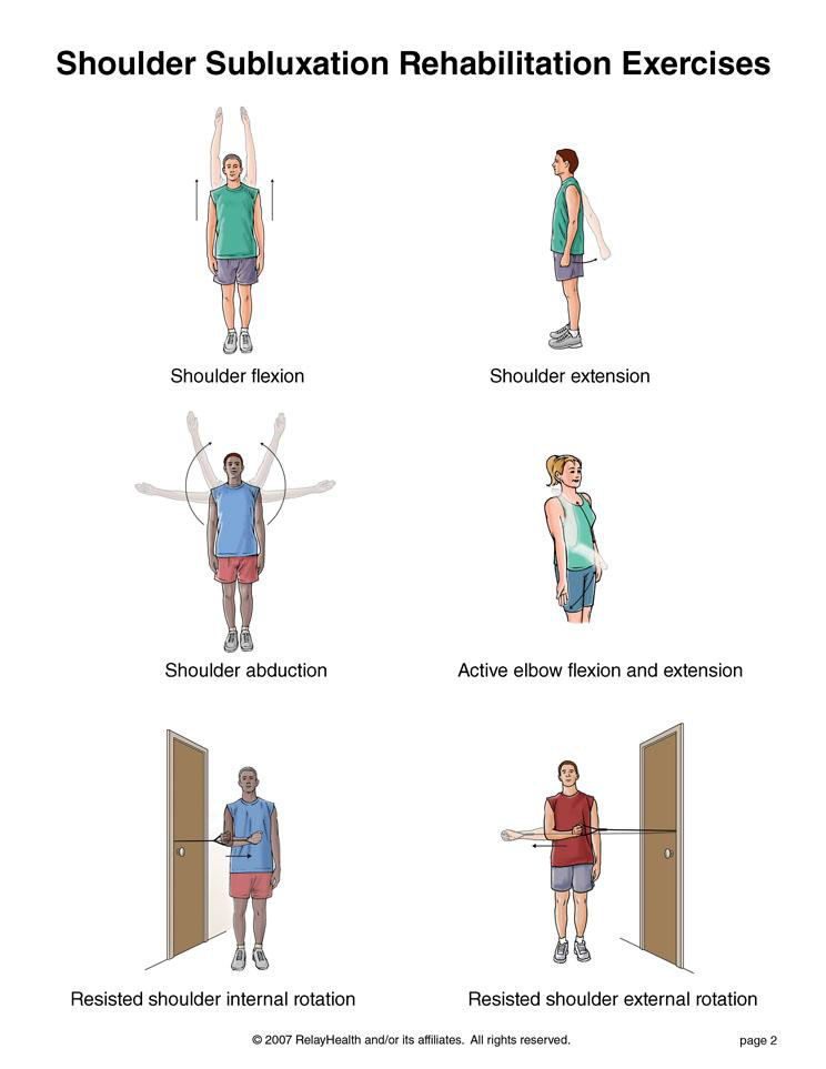
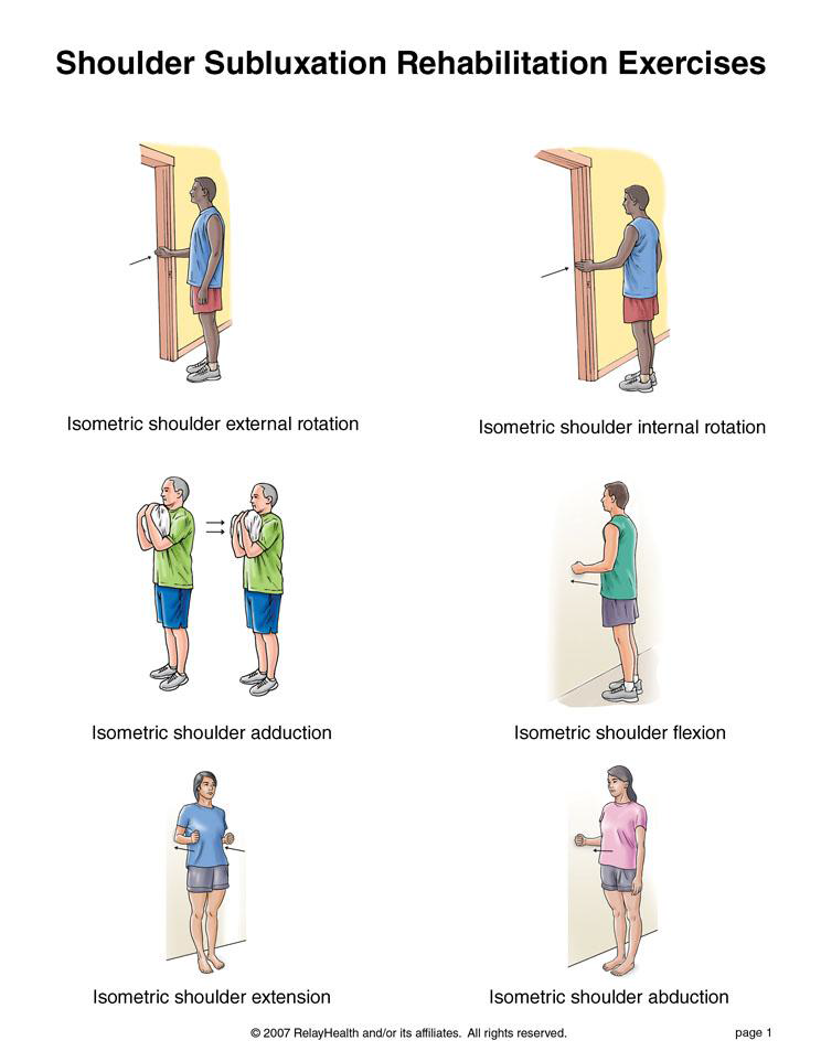
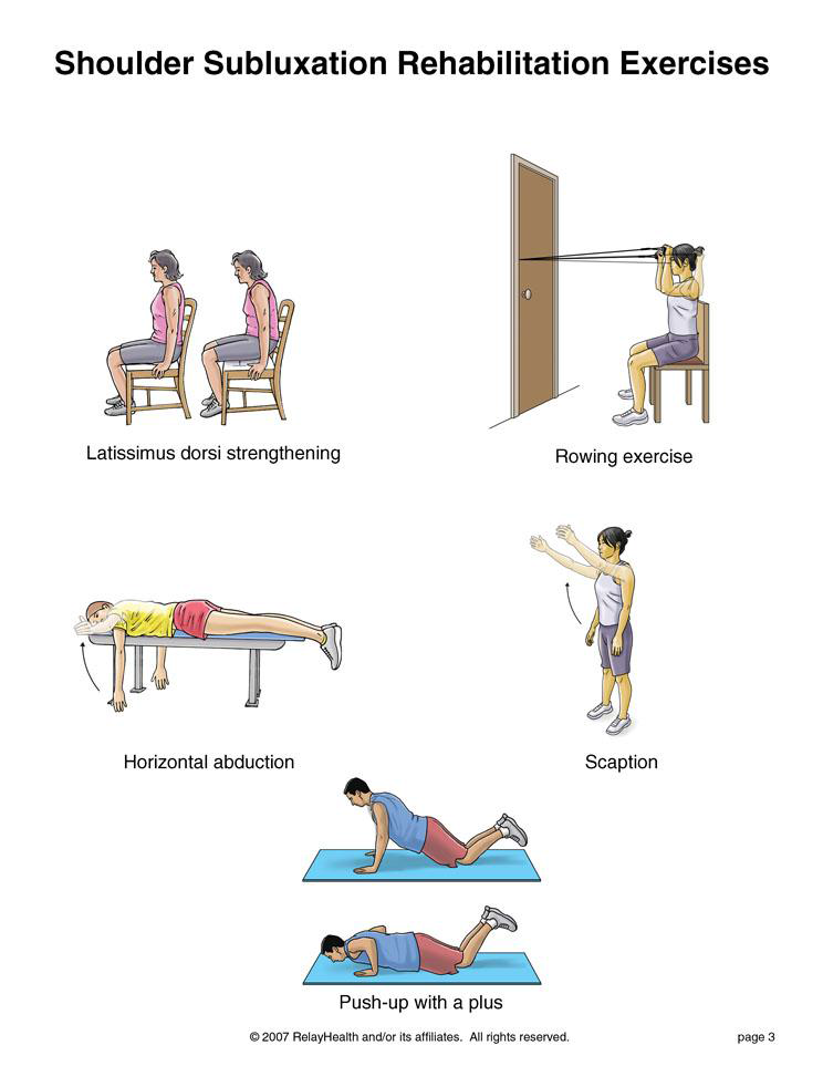
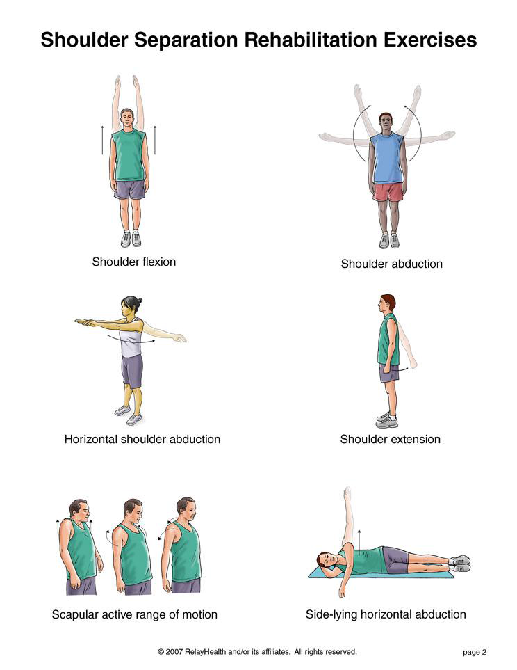
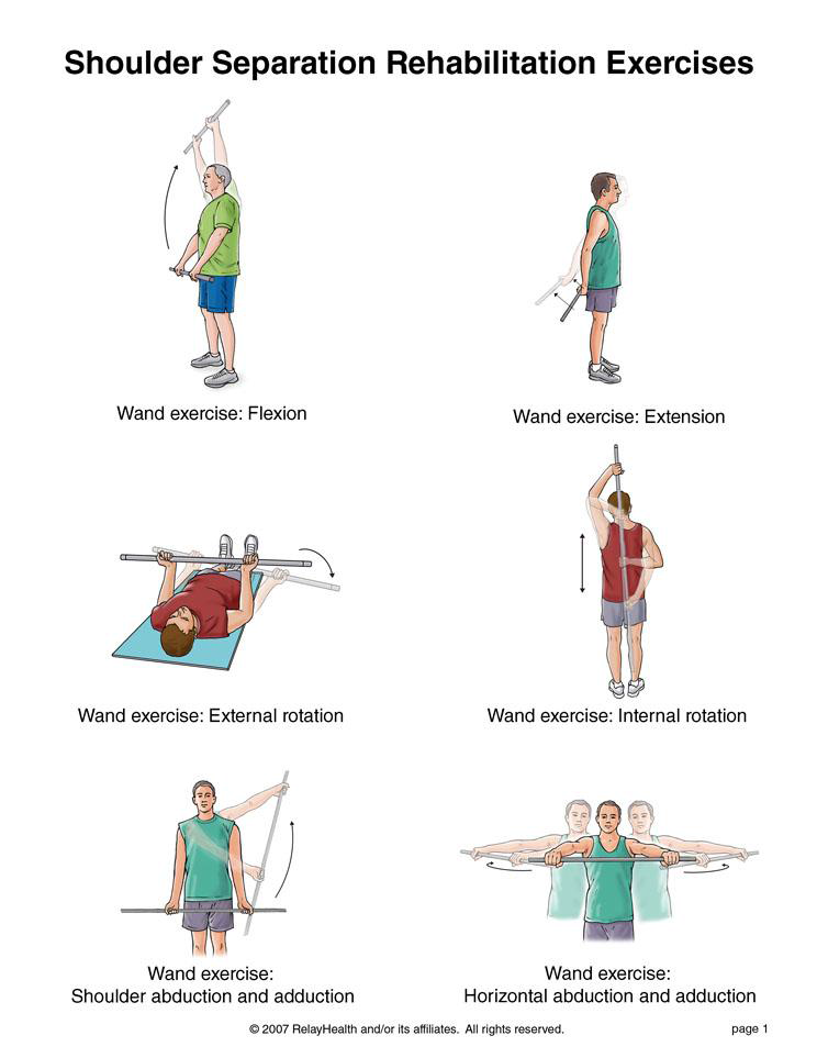
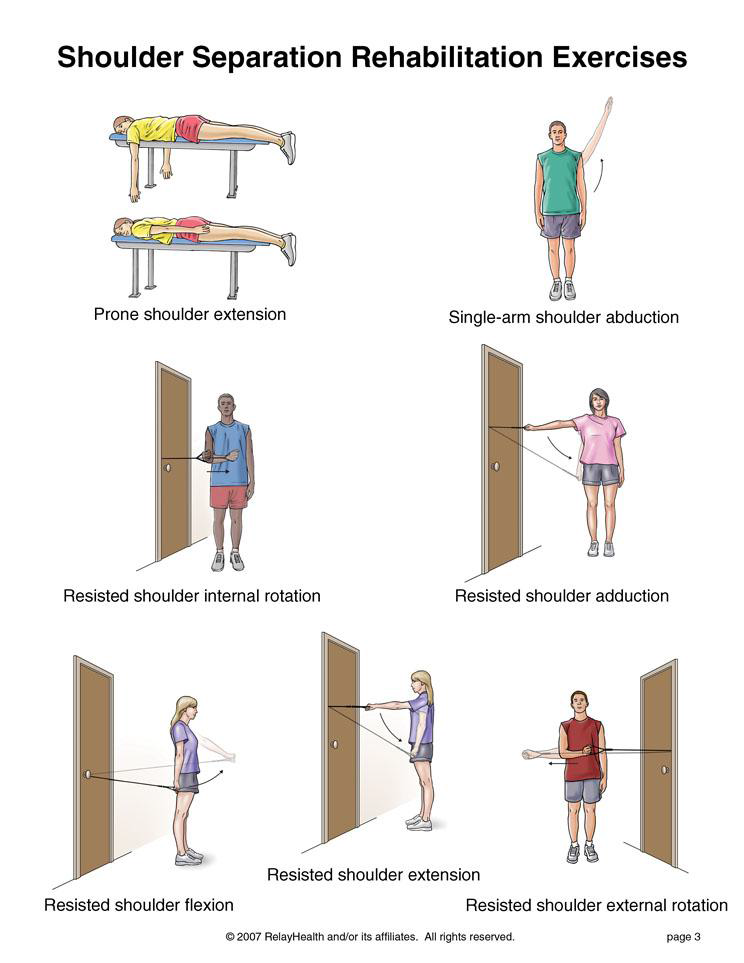
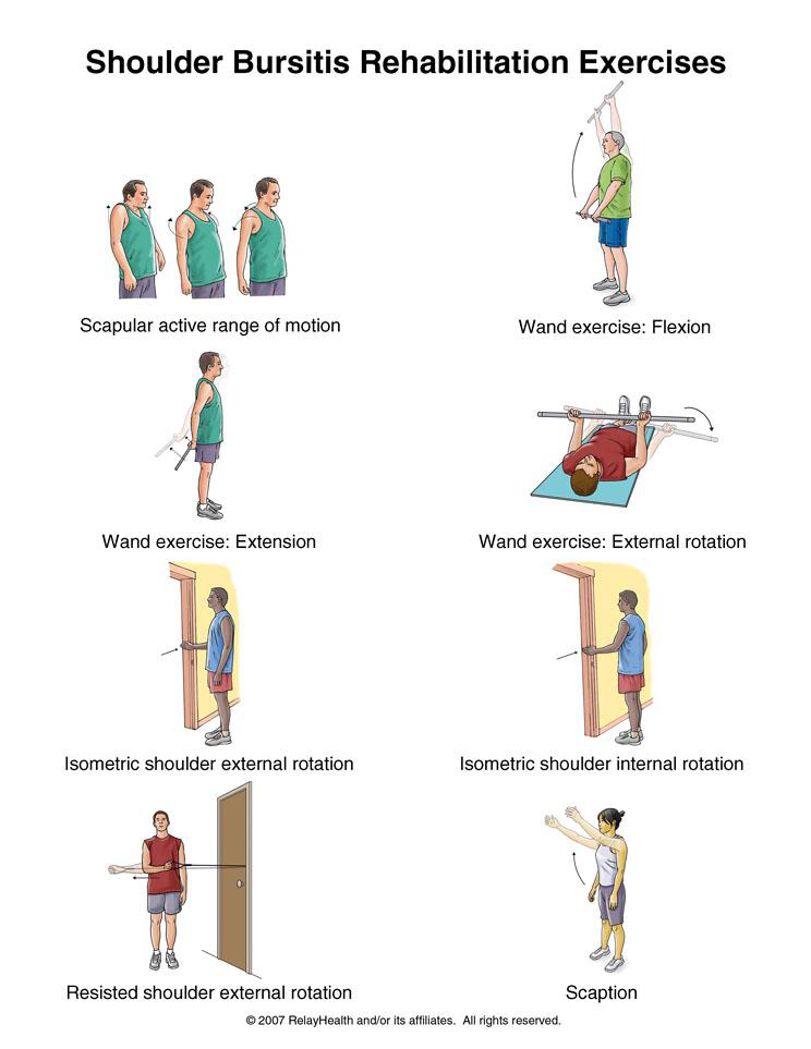
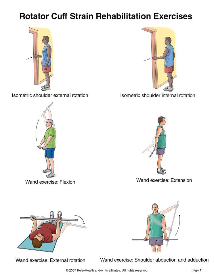
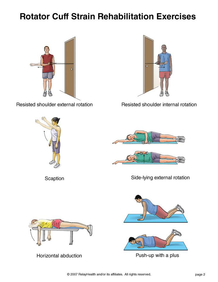
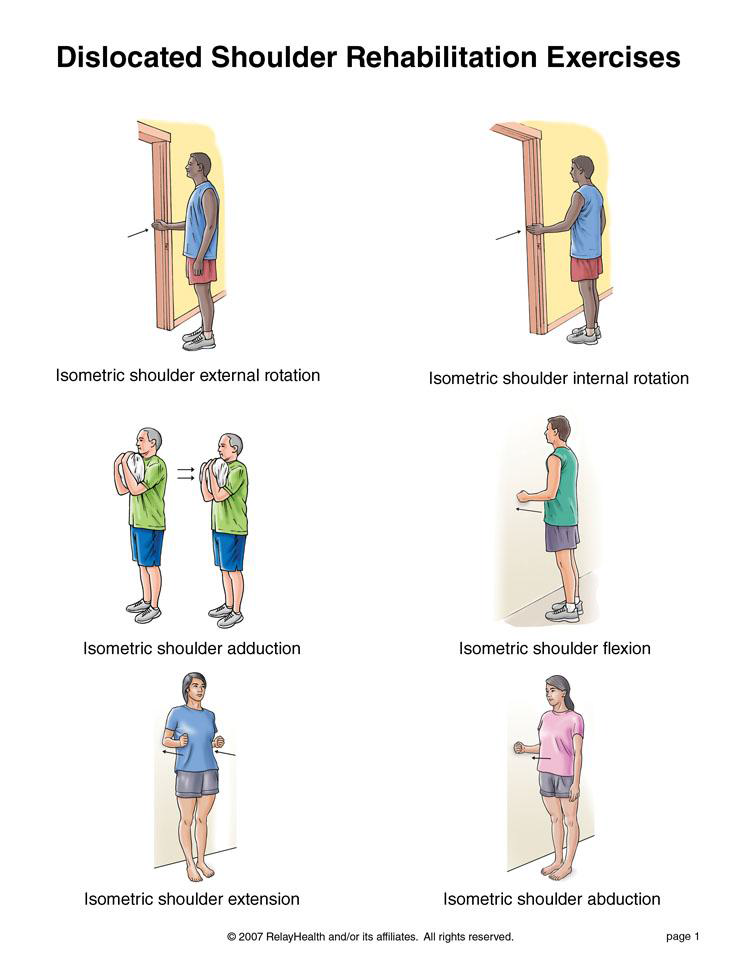
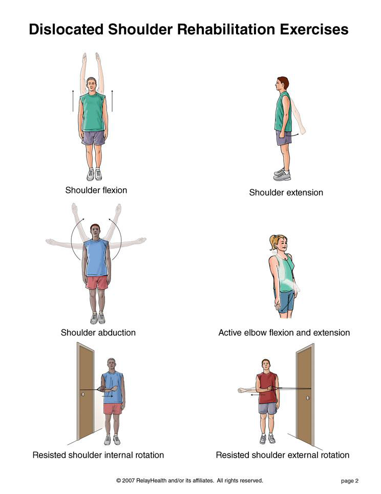
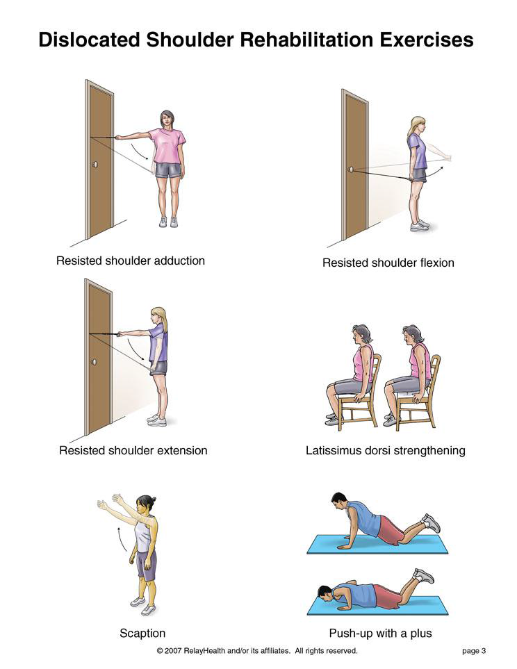
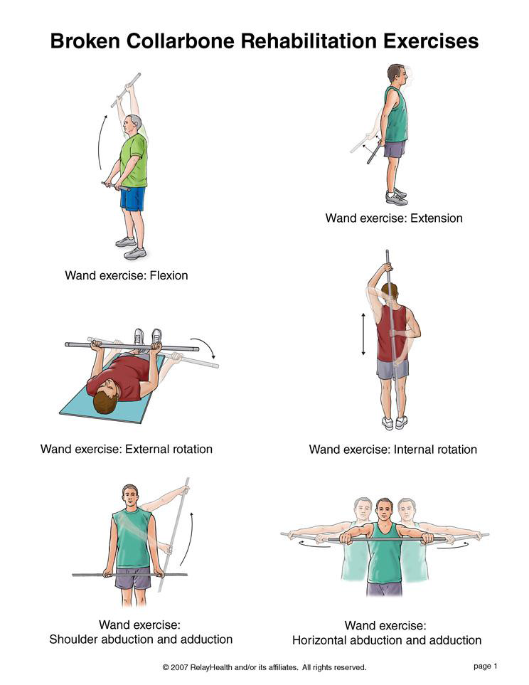
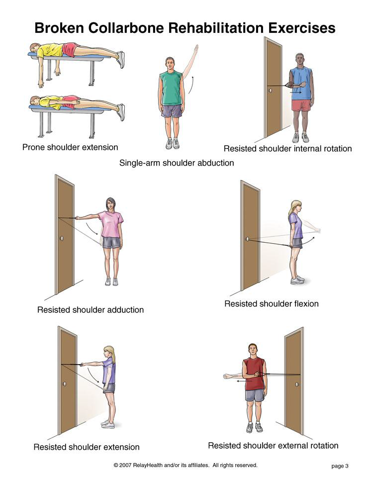
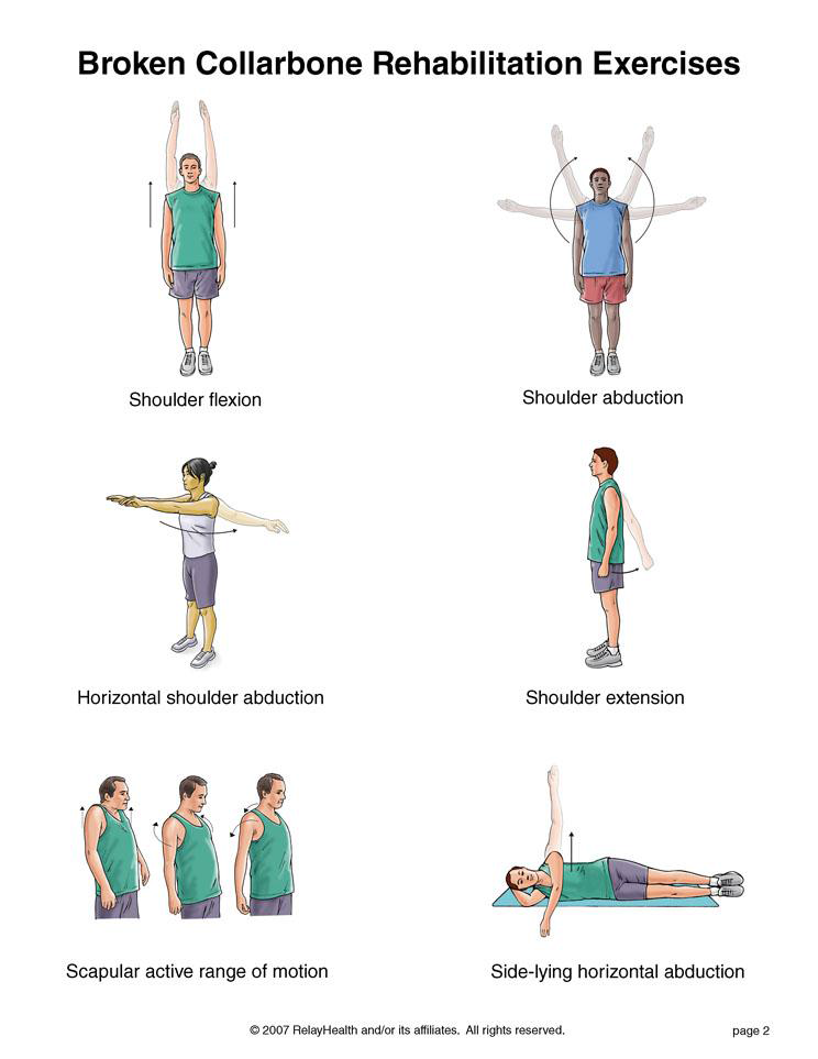
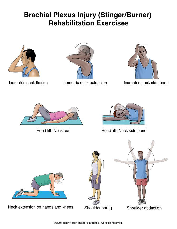
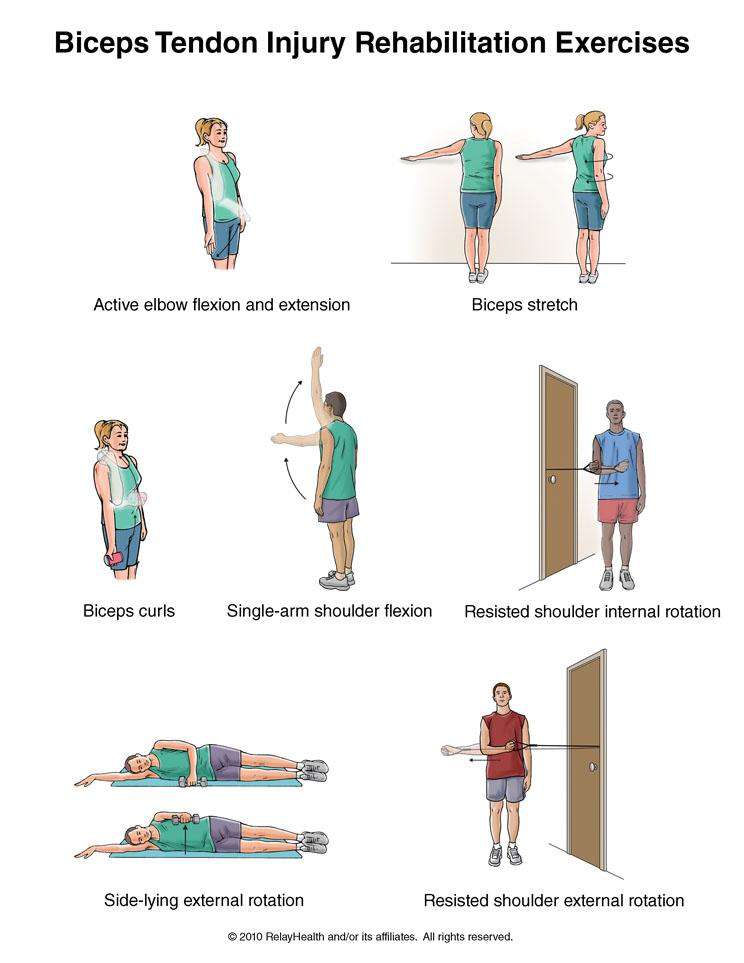
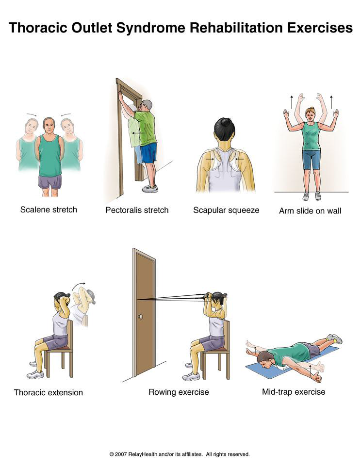
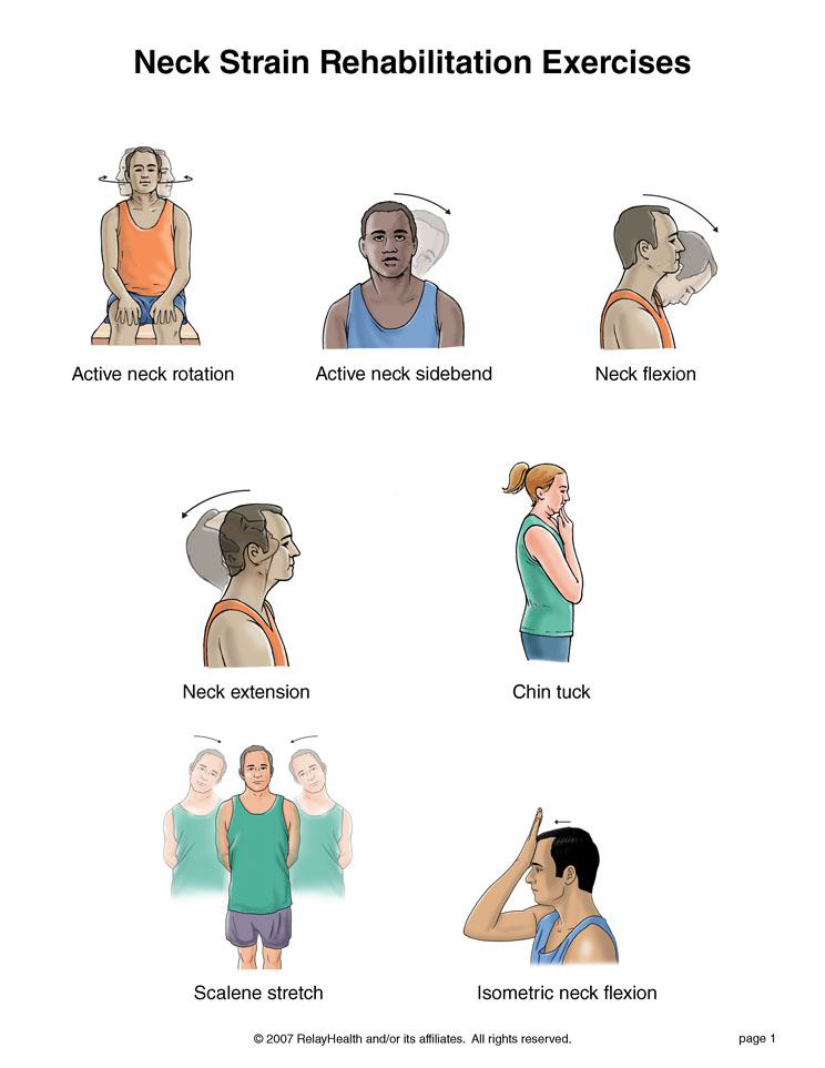
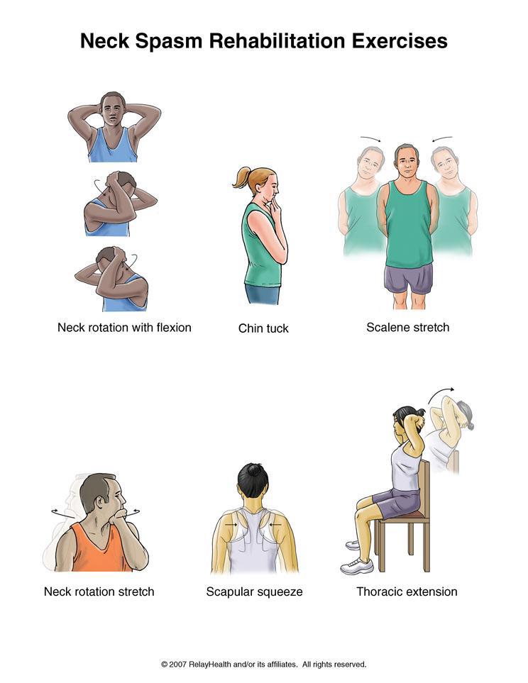
Physical Therapy:
HealthZone MD Family and Sports Medicine Wellness Center takes a comprehensive approach to sports injury management, leveraging the expertise of both sports medicine physicians and physical therapists to provide optimal care for athletes. Here’s how our physicians collaborate with physical therapists and utilize rehabilitation exercises to improve sports injury management:
- Comprehensive Assessment:
- Our sports medicine physicians conduct thorough assessments of athletes’ injuries, including physical examinations, diagnostic imaging (such as X-rays, MRI, or ultrasound), and functional movement assessments.
- Based on the assessment findings, the physician collaborates with physical therapists to develop individualized treatment plans tailored to the athlete’s specific injury, functional limitations, and athletic goals.
- Treatment Planning and Coordination:
- Physicians work closely with physical therapists to coordinate treatment plans that integrate medical interventions with rehabilitation strategies.
- Treatment plans may include a combination of medical interventions (such as medications, injections, or bracing) and physical therapy modalities (such as therapeutic exercises, manual therapy, and modalities like ultrasound or electrical stimulation).
- Prescription of Rehabilitation Exercises:
- Our sports medicine physicians prescribe rehabilitation exercises designed to address the underlying causes of the injury, improve strength, flexibility, balance, proprioception, and functional movement patterns.
- Physical therapists collaborate with physicians to ensure that the prescribed exercises are safe, effective, and aligned with the athlete’s stage of recovery.
- Supervision and Progress Monitoring:
- Physical therapists oversee the implementation of rehabilitation exercises, providing hands-on guidance, instruction, and feedback to athletes to ensure proper technique and adherence to the prescribed program.
- Our physicians collaborate with physical therapists to monitor athletes’ progress throughout the rehabilitation process, adjusting treatment plans as needed based on objective measures of improvement and subjective feedback from the athlete.
- Gradual Return-to-Sport Progression:
- Physicians and physical therapists work together to develop structured return-to-sport protocols that safely reintroduce athletes to their sport-specific activities.
- Rehabilitation exercises are incorporated into the return-to-sport progression to gradually increase the intensity, volume, and complexity of training while minimizing the risk of reinjury.
- Education and Injury Prevention:
- Our sports medicine team educates athletes on proper body mechanics, injury prevention strategies, warm-up and cool-down routines, and lifestyle modifications to reduce the risk of future injuries.
- Physical therapists teach athletes how to perform rehabilitation exercises correctly, provide guidance on home exercise programs, and offer ergonomic advice to optimize performance and prevent overuse injuries.
- Ongoing Collaboration and Communication:
- Collaboration between physicians and physical therapists is ongoing throughout the rehabilitation process, with regular communication to discuss progress, challenges, and adjustments to the treatment plan.
- Our sports medicine team works together seamlessly to provide cohesive, integrated care focused on maximizing athletes’ recovery, restoring function, and promoting long-term musculoskeletal health and athletic performance.
By leveraging the expertise of both sports medicine physicians and physical therapists, HealthZone MD Family and Sports Medicine Wellness Center delivers comprehensive, multidisciplinary care that optimizes sports injury management and facilitates athletes’ return to peak performance safely and effectively.
Xray Facility:
At HealthZone MD Family and Sports Medicine Wellness Center, we leverage state-of-the-art digital x-ray radiography technology to diagnose, treat, and manage sports-related injuries and fractures with precision and efficiency. We utilize this advanced imaging technology in our practice for :
Accurate Diagnosis:
Digital x-ray radiography allows our sports medicine physicians to obtain high-quality images of bones, joints, and soft tissues quickly and accurately.
These images enable us to visualize and assess the extent of sports-related injuries, such as fractures, dislocations, sprains, strains, and joint abnormalities.
With digital x-rays, we can zoom in on specific areas of interest, adjust image contrast and brightness, and manipulate images to enhance diagnostic clarity, leading to more accurate and confident diagnoses.
Timely Treatment:
Rapid image acquisition and processing with digital x-ray technology expedite the diagnostic process, allowing our physicians to promptly assess and diagnose sports-related injuries.
Timely diagnosis enables us to initiate appropriate treatment interventions promptly, minimizing delays in care and optimizing outcomes for athletes.
For acute fractures and traumatic injuries, digital x-rays help guide immediate treatment decisions, such as immobilization, splinting, or referral for surgical intervention, as needed.
Comprehensive Treatment Planning:
Digital x-ray images serve as valuable tools for treatment planning, allowing our physicians to develop personalized treatment plans tailored to the unique needs and goals of each athlete.
With detailed anatomical information provided by digital x-rays, we can accurately localize injuries, assess fracture stability, and determine the most appropriate course of action, whether conservative management, orthopedic intervention, or rehabilitation.
Monitoring and Follow-up:
Digital x-ray radiography facilitates ongoing monitoring of sports-related injuries and fractures throughout the recovery process.
Repeat imaging with digital x-rays enables our physicians to assess healing progress, monitor fracture alignment, and evaluate treatment efficacy over time.
Regular follow-up x-rays allow us to make informed decisions regarding treatment modifications, rehabilitation progress, and return-to-sport readiness based on objective evidence of healing and recovery.
Patient Education and Engagement:
Digital x-ray images provide visual evidence of sports-related injuries, enhancing patient understanding of their condition and treatment options.
Our physicians use digital x-rays as educational tools to explain injury mechanisms, treatment recommendations, and expected outcomes to athletes and their families, empowering them to actively participate in their care decisions.
By involving patients in the diagnostic and treatment process, we foster greater engagement, compliance, and satisfaction with the treatment plan, leading to better overall outcomes.
Overall, digital x-ray radiography plays a pivotal role in the comprehensive management of sports-related injuries and fractures at HealthZone MD Family and Sports Medicine Wellness Center, enabling accurate diagnosis, timely treatment, personalized care, ongoing monitoring, and patient education. Through the integration of advanced imaging technology into our practice, we strive to deliver the highest quality of care and optimize outcomes for athletes of all levels.